Superior Oblique Tendon Inferior Oblique Muscle Art Labeling Activity
We have a lot of muscles in our bodies (literally, over 600). Muscles let u.s. to move and part. In general, they work in pairs. Usually as one muscle contracts (or shortens), the opposing muscle (known every bit the antagonist) elongates and vice versa. For example, recall about when you curve your arm to bring nutrient to your mouth. Multiple muscles on the front end of your arm shorten (biceps, brachialis, etc.) to permit for this to happen. Conversely, as you do this, the antagonist muscle (triceps) elongates. Then when yous demand to straighten your arm out, the triceps will shorten and the biceps (and others) will elongate.
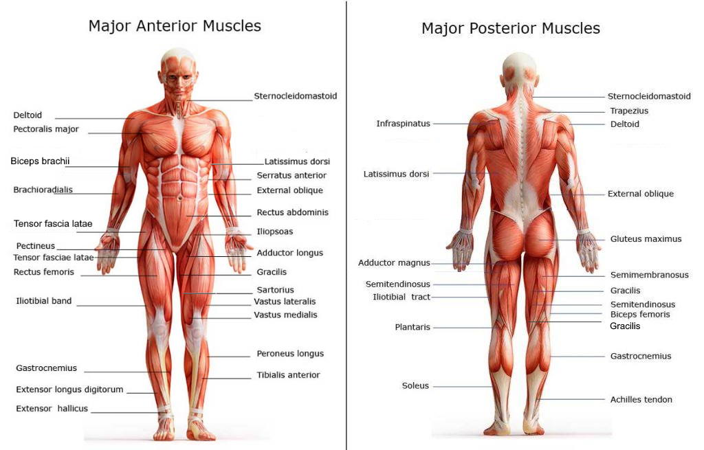
In this article, nosotros introduce to y'all the major muscles on the front of the body.
Anatomic Terms
To better understand muscles and how they piece of work, it's important to briefly familiarize ourselves with anatomical terms. Anatomical terms allow wellness care professionals to accurately communicate to others which part of the trunk may be afflicted by disorder or a affliction. Ultimately, communicating using anatomical terms makes information technology like shooting fish in a barrel to communicate descriptions of body areas regardless of the individual'southward position. For example, suppose a doc was trying to draw an area of the body to another physician on a patient who is lying face up down? Anatomical terms would allow this discussion to happen with ease.
Planes are ofttimes used to describe location of structures or to describe directionality of motion. A plane is a theoretical line that divides the body. Oft, these terms are used within the context of advanced medical imaging studies such every bit computed tomography (CT) and magnetic resonance imaging (MRI) scans.
You lot can see that the three bones planes (sagittal, coronal, transverse) intersect one another at correct angles. For "normal" human bodies, the right and left sides are mirror images if divided correct downward the center by thesagittalplane every bit shown in the image below.
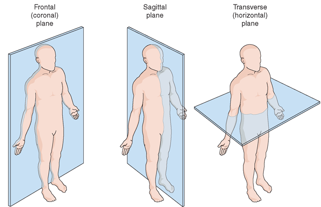
Motion about these planes can be described past anaxis of movement. For example, motion almost the sagittal axis occurs in the sagittal plane e.g. bending forwards at the waist (known as flexion) and backwards (known as extension). Accordingly, move about the transverse axis occurs in the transverse aeroplane e.yard. twisting at the waist (known equally rotation). Finally, movement well-nigh the coronal axis occurs in the coronal plane eastward.g. bending your body to the left or correct.
Prior to reading on, y'all should review anatomical terms. Additionally, you should review the glossary of terms at the end of this article.
List of Major Inductive Muscles

Adductor longus
A musculus of the medial thigh that originates on the pubis. It inserts onto the linea aspera of the femur. It adducts, flexes, and rotates the thigh medially. It is controlled past the obturator nerve. It pulls the leg toward the body's midline (i.e. adduction)
Biceps brachii
An upper arm muscle composed of two parts, a long head and a brusk head. This musculus flexes the elbow and shoulder as well as supinates the forearm (i.eastward. rotates the forearm so the palm is facing the ceiling). The long head originates just above the shoulder socket on the scapula and blends with the short head onto the radius bone of the forearm. The short caput originates on the coracoid process of the scapula.
Brachioradialis
A muscle lying on the lateral side of the forearm. This muscle connects the humerus to the radius at the styloid procedure. It flexes the forearm. Likewise depending on the position of your hand, information technology can rotate the forearm in either direction.
Coracobrachialis
The coracobrachialis is the smallest of the three muscles that attach to the coracoid process of the scapula. (The other ii muscles that attach here are the pectoralis minor and the short head of the biceps brachii.) Information technology is situated at the upper and medial part of the arm. It is supplied past the musculocutaneous nerve. The coracobrachialis draws the humerus forward (shoulder flexion) and towards the trunk (shoulder adduction) at the shoulder (glenohumeral) joint.
Deltoid
This large triangular muscle wraps around the shoulder joint and connects the scapula, clavicle (collar-bone) and humerus. It is a 3-part muscle with anterior (front end), middle, and posterior (back) heads. It is controlled past the axillary nerve. The front fibers flex the arm and the middle fibers help housebreak the arm (bring the arm abroad from the body). Posterior (back) fibers assistance to extend the arm.
Extensor Hallucis Longus (EHL)
The extensor hallucis longus or EHL is a thin muscle situated between the tibialis inductive and the extensor digitorum longus (EDL) that mainly functions to extend the great toe (bring it towards the ceiling). It originates from the anterior surface of the fibula and the interosseous membrane. It is supplied by the deep peroneal nerve.
Extensor Digitorum Longus (EDL)
This muscle arises from the lateral condyle of the tibia. The muscle passes over the ankle under a fibrous sheath called the extensor retinaculum and divides into four separate tendons. These tendons run along the superlative of the foot and insert into the four lesser toes. This muscle allows united states of america to extend our toes and our human foot (an action known as dorsiflexion)
External oblique muscle
This is a diagonally oriented muscle that helps to tighten the abdomen. It's the largest and the virtually outermost of our three intestinal muscles. It has limited deportment in both flexion and rotation of the vertebral column. 1 side of the obliques contracting can create lateral flexion. It also helps in pulling in the belly. The two muscles on either side of the chest come together to course a gristly sail. These muscles assist the rectus abdominis to go on the abdominal organs in place.
Gastrocnemius
The large musculus of the posterior role of the lower leg. Information technology is the about superficial of the calf muscles. The gastrocnemius has 2 heads, 1 originating along the outside of the caput and condyle of the femur and the other originating along the medial popliteal surface of the femur. Both heads attach to the dorsum surface of the calcaneus, also called the heel bone the heel with the calcaneal tendon, also called the Achilles. When it contracts the gastrocnemius plantar flexes the talocrural joint bending the foot downward, flexes the articulatio genus, and allows a person to stand up on tip toes. It is innervated by the tibial nerve. It's proper name means "belly of the leg."
Gluteus medius
A musculus of the hip originating on the lateral surface of the ileum and inserted in the greater trochanter of the femur. Information technology abducts and medially rotates the past and is controlled by the superior gluteal nerve.
Gracilis
A long slender musculus on the medial attribute of the thigh.
Iliopsoas
The compound iliacus and psoas magnus muscles.
Iliotibial band (ITB)
– a dense band of non-contractile tissue, called fascia, that covers the gluteal region and into this tensor fascia lata of an gluteus maximus are attached. Distallly the ITB inserts into the kneecap, tibia and fibula caput.
Latissimus dorsi
A back muscle that pulls the arm down and dorsum. It is responsible for extension,adduction, and (medial) internal rotation of the shoulder articulation. Information technology also helps in extension and lateral flexion of the lumbar spine. The name ways "widest of the back." This muscle supports the arm when it is moved above the caput. If yous button your arm hard confronting your side, you will feel this muscle tighten up.
Pectineus
Pectoralis major
A chest musculus that pulls the arm in towards the body. This is one of the internal rotator muscles that attach the humerus and internally rotate the arm. The pectoralis major originates forth the clavicle, downward the sternum, and across the ribs and inserts into the humerus. This muscle tin contribute to excessive internal rotation of the arm or scapular abduction.
Peroneus longus
Fibularis longus muscle. A muscle forth the outside of the leg that bends the foot out at the ankle. The fibularis longus originates from the head and upper lateral surface of the fibula, runs in a bony groove forth the bottom of the pes to adhere on the other side at the base of the first metatarsal and the neighboring medial cunieform bone, and acts to evert the foot; it is innervated past the superficial fibular nervus.
Rectus abdominis
Aalso known every bit the "six pack", is a paired musculus running vertically on each side of the front wall of the abdomen. There are two parallel muscles, separated by a midline ring of connective tissue chosen the linea alba. The rectus abdominis is an of import postural musculus. It is responsible for pulling the rib cage toward the pelvis. The rectus abdominis helps when we exhale while breathing and forcefully exhaling. It likewise helps in keeping the internal organs intact and in creating pressure within the abdomen, such as when exercising or lifting heavy weights, during forceful defecation or pushing during childbirth.
Rectus Femoris
a musculus of the anterior thigh originating on the iliac spine and upper margin of the acetabulum and inserted in the tibial tuberosity by style of the patellar ligament. It extends the leg, contributes to flexion of the thigh, and is controlled past the femoral nerve.
Sartorius
A long, ribbon-shaped musculus in the leg that flexes, abducts, laterally rotates the thigh, and flexes the lower leg. This musculus, the longest in the trunk, enables the crossing of the legs in the tailors'due south position, the function for which information technology is named. Information technology is strapped shaped and winds across the front of the thigh, from the hip to the inner side of the tibia. When information technology contracts it bends and rotates the thigh.
Serratus anterior
This muscle is divided into 3 named parts: serratus anterior superior, serratus anterior intermediate, serratus anterior inferior and runs from the front of the chest around the side to the scapula. The anterior serratus pulls the scapula outward which lifts the shoulder. It keeps the scapula in position close to the chest wall, abducts the scapula, and turns information technology upward to raise the bespeak of the shoulder. If the scapula is stock-still, the serratus inductive can elevate the ribs. The serratus anterior is controlled by the long thoracic nervus. Serratus ways "saw-shaped" and describes this muscle's jagged shape.
Sternocleidomastoid
is a paired muscle in the superficial layers of the front role of the neck. Information technology tilts the head to its own side and rotates the caput and so the head faces the opposite side. Information technology is also an accessory muscle of breathing out and raises the sternum.
Tensor fasciae lata (TFL)
originates on the anterior portion of the iliac crest and ASIS and inserts into the ITB. Information technology flexes, medially rotates, and abducts the leg and can cause pelvic rotation problems.
Teres major muscle
is a muscle of the arm and ane of six scapulohumeral muscles. It is not part of the rotator cuff. The teres major is a medial rotator and adductor of the humerus and assists the latissimus dorsi in cartoon the previously raised humerus down and backward (extension, but not hyper extension). Information technology as well helps stabilize the humeral head in the glenoid crenel.
Tibialis anterior
An extensor musculus that straightens or lifts the foot. A muscle of the leg originating on the lateral condyle of the tibia and the interosseus membrane betwixt the tibia and the fibula and inserted in the first cunieform and first metatarsal bones. Information technology dorsi flexes and inverts the foot, supports the arch, and is controlled by the deep peroneal nerve.
Vastus lateralis
A muscle of the anterior thigh originating on the linea aspera and the greater trochanter of the femur and inserted in the tibial tuberosity by style of the patellar ligament. It extends the leg and is controlled by the femoral nerve.
Vastus medialis
a muscle of the inductive thigh originating on the linear aspera and inter-trochanteric line of the femur and inserted in the tibial tuberosity via the patellar ligament. It extends the leg and is controlled by the femoral nervus.
Glossary
ASIS
anterior superior iliac spine.
Distal
Farther from the midline. For instnace, the wrist is distal to the elbow joint – that is, the wrist is farther away from the midline than the elbow.
Proximal
Closer to the midline, that is – the elbow is distal to the wrist.
Adduction
Movements that bring limbs towards the midline. For instance, bringing your arm down to your from sideward extension is adduction.
Abduction
To abduct a limb or to movement it away from the midline. Lifting the arms sidewards from resting position is an example of abduction.
Extension
Movements that straighten out a joint, for instance extending the human knee involves straightening the knee joint.
Flexion
Shortening of a muscle. Flexion will oftentimes curve a limb, such as when flexing the bicep, thus angle the elbow.
Rotation
Motion that occurs in the horizontal plane. For instance, when the arms are at residual at a person's sides, external and internal rotation will supinate or pronate the easily and forearms.
Insertion
The distal attachment of a musculus. The insertion is the segment that moves during muscle flexion. For case, the bicep inserts forth the radial tuberosity. When the bicep is flexed, the radius or forearm moves towards the upper arm.
Origin
The proximal attachment of the muscle, ofttimes considered the anchor of movement. For instance, the bicep originates from the scapula and shoulder. Thus whatsoever motility performed by the biceps will bring the insertion closer to the origin.
Innervation
Nerve supply of a muscle. Unlike nerves branch out throughout the body to provide each musculus electrical impulses from the brain to trigger movement.
nuneztolopead1963.blogspot.com
Source: https://www.healthpages.org/health-a-z/anatomy-major-anterior-muscles/
0 Response to "Superior Oblique Tendon Inferior Oblique Muscle Art Labeling Activity"
Enregistrer un commentaire